Advanced Microscopy and Imaging
उन्नत माइक्रोस्कोपी और इमेजिंग के बारे में
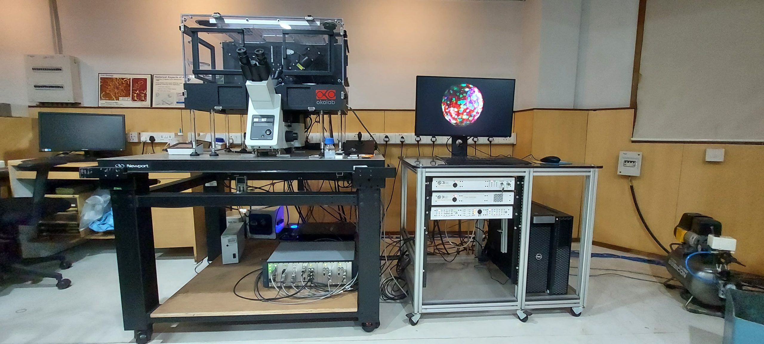
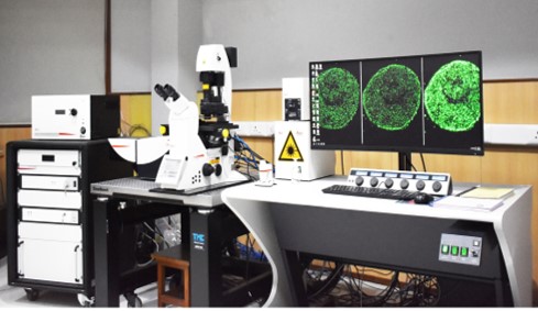
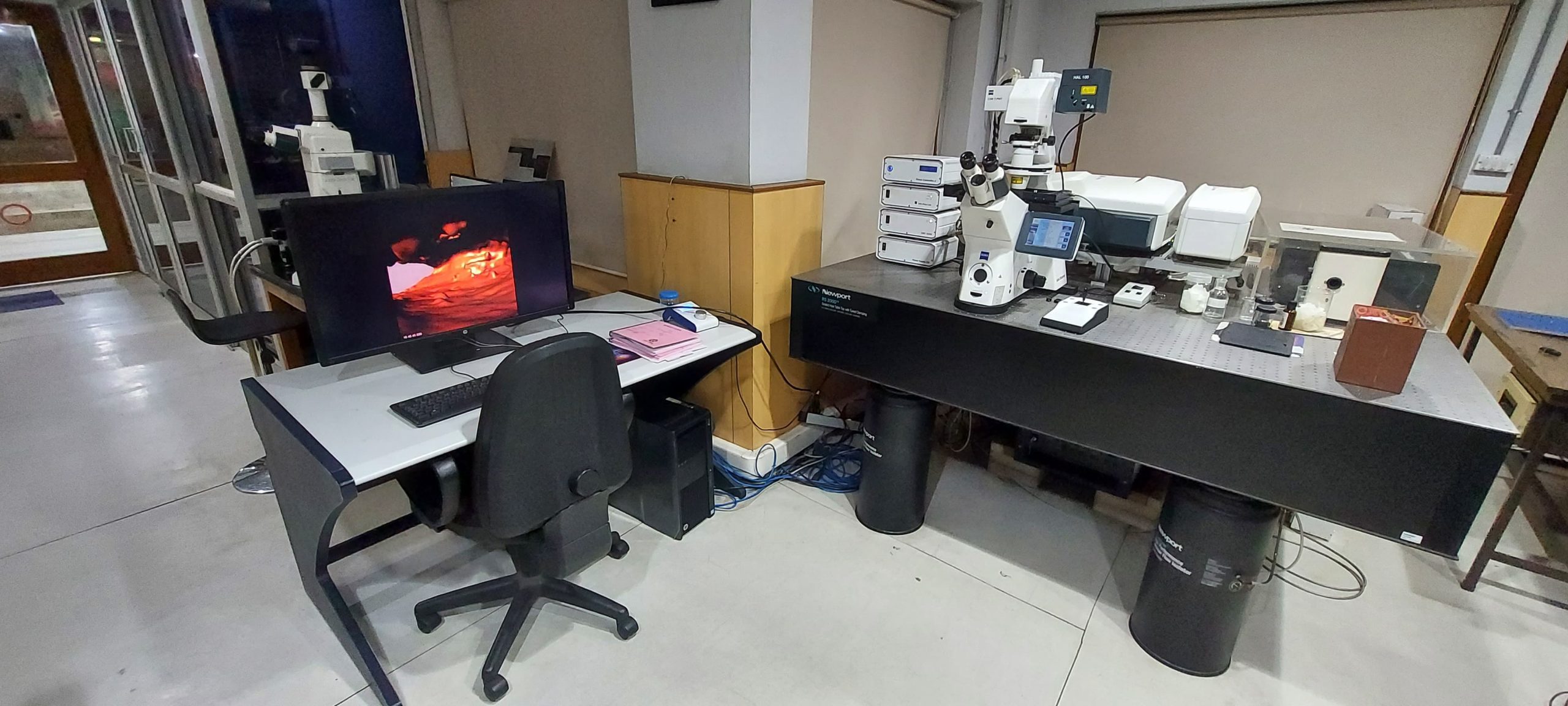
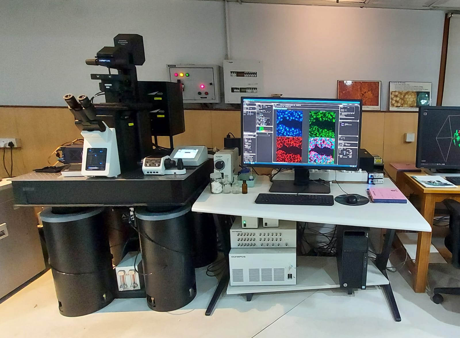
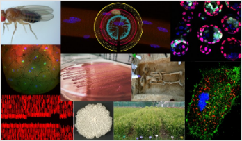
कोशिकीय एवं आणविक जीव विज्ञान केंद्र (CCMB) में उन्नत माइक्रोस्कोपी और इमेजिंग सुविधा (AMIF) की स्थापना देश के पहले लेजर स्कैनिंग कॉन्फोकल माइक्रोस्कोपों में से एक के साथ की गई थी। हमारी अत्याधुनिक सुविधा जैविक इमेजिंग तकनीकों की एक विस्तृत श्रृंखला प्रदान करती है, जिसमें कॉन्फोकल और मल्टी-फोटॉन माइक्रोस्कोप, एक प्रतिदीप्ति सहसंबंध स्पेक्ट्रोस्कोप (fluorescence correlation spectroscope), परमाणु बल माइक्रोस्कोप (atomic force microscope), सुपर-रिज़ॉल्यूशन माइक्रोस्कोप, रमन माइक्रोस्कोप, लाइट शीट माइक्रोस्कोप, एक लाइव सेल इमेजिंग सिस्टम, TIRF माइक्रोस्कोप, और डिकॉन्वोल्यूशन सॉफ्टवेयर, हाई-स्पीड और 3D स्टीरियो इमेजिंग क्षमताओं वाले विभिन्न अपराइट और इनवर्टेड प्रतिदीप्ति माइक्रोस्कोप शामिल हैं। यह सुविधा सीसीएमबी शोधकर्ताओं और बाहरी उपयोगकर्ताओं, दोनों के लिए खुली है। आप हमें सीसीएमबी मुख्य परिसर के कमरा नंबर C-104 में पा सकते हैं। उपकरणों की विस्तृत जानकारी के लिए, कृपया ‘उपकरण’ (Equipment) टैब देखें।
Venkata Mahesh Nitla, PhD
Imaging ScientistDr. Mahesh Nitla received his PhD in Biology (Protistology) from University of Pisa, Italy.
Google Scholar

Nandini Rangaraj
Imaging ScientistGoogle Scholar

Mr. Suman Bandari
Imaging specialistGoogle Scholar
Equipments
AxioZoom V16 Zoom Microscope
3i Marianas LightSheet
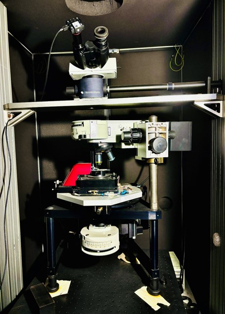
AFM-Raman integrated system
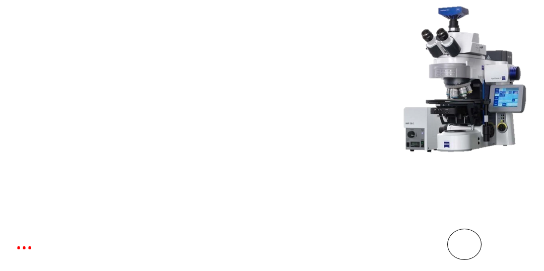
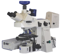
Axio Imager Z2 with Apotome
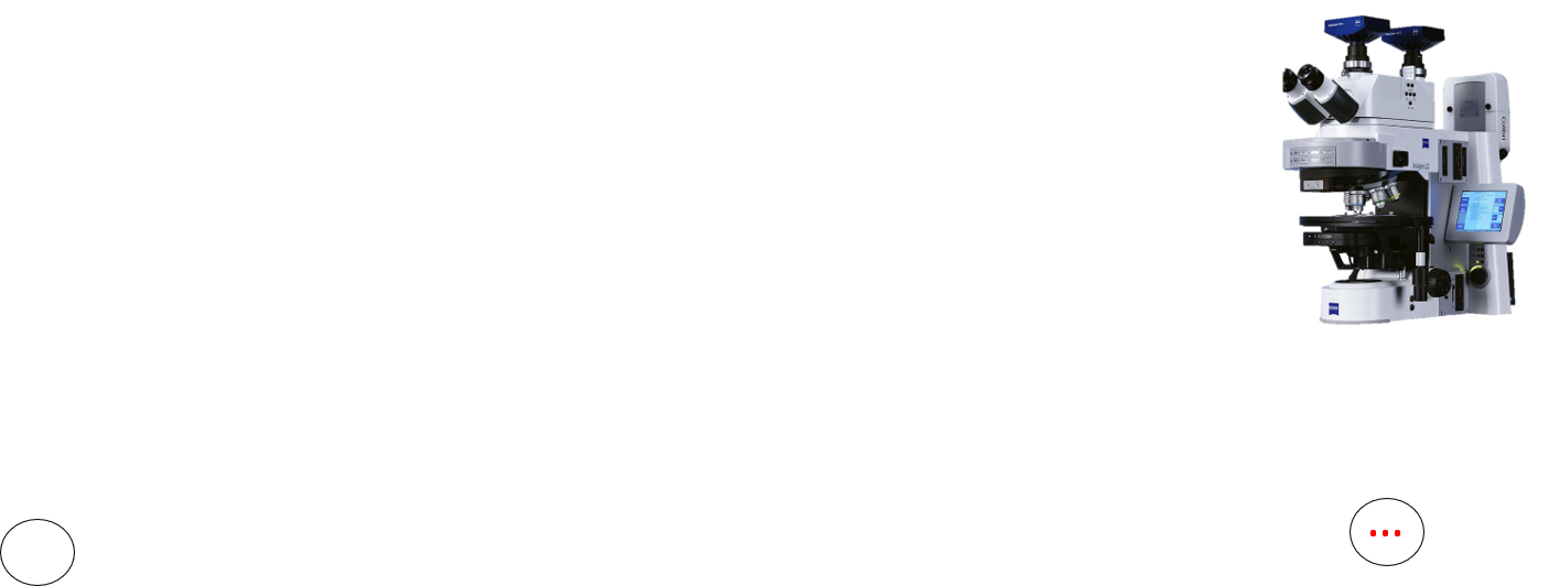
Axio Imager Z2
Axio Zoom.V16
Leica 3x STED
LSM 880
Olympus FV3000
Training Program for In-House Users: Handling Sophisticated Imaging Systems
1. Access Control & Training (In-house users)
- Lecture (2 hours):
- Basics of microscopy
- Fluorescence microscopy
- Digital imaging techniques
2. System Demonstrations
- Basics of Microscopy and Fluorescence Microscopy (2.5 hours):
- Transmitted light microscopy techniques: Bright field, DIC, Phase contrast,
Darkfield - Kohler illumination
- Fluorescence microscopy
- Hands-on: Operate Leica DM6 fluorescence microscope and complete
assignment - Confocal Microscopy (2 hours):
- Demonstration of Leica SP8 / Zeiss LSM 880 confocal microscope
- Overview of confocal imaging principles and setup
3. Examination
- Online MCQ Test (45 minutes):
- Complete multiple-choice questionnaire on microscopy concepts and
techniques - Instant results upon submission
4. Confocal Hands-on Exercise (2-3 hours)
- Practical Session:
- Users complete assignment using sub-resolution beads on the Leica SP8
Confocal/ LSM 880 confocal microscope - Independent operation to apply learned concepts
5. Submission of Assignments
- Submit assignment results after completing the hands-on exercise
6. Facility Access
- Facility Access: Gain access to the imaging facility after assessment by the
facility in charge
This structured, point-wise program ensures that users receive comprehensive training,
theoretical knowledge, and hands-on practice to confidently operate imaging systems.
Training Programs/SDP
The Advanced Microscopy and Imaging Facility (AMIF) conducts an annual microscopy
course, offering hands-on training in cutting-edge imaging techniques. This intensive
program covers confocal, light-sheet, and super-resolution microscopy, with expert-led
sessions on sample preparation, image acquisition, and analysis. Designed for researchers
and students, the course provides a unique opportunity to explore the latest advancements
in microscopy and enhance imaging skills for biological research.
No safety button available
Resources
A number of genetic disorders are known to result from the defects in a single gene. Although rare in comparison to infectious diseases, genetic disorders cause enormous misery as many of them do not have a permanent cure. In the absence of specific treatment, molecular diagnosis, carrier detection and prenatal diagnosis or pre-implantation genetic diagnosis for these disorders with appropriate genetic counseling is the best approach to prevent their recurrence/transmission.
A number of genetic disorders are known to result from the defects in a single gene. Although rare in comparison to infectious diseases, genetic disorders cause enormous misery as many of them do not have a permanent cure. In the absence of specific treatment, molecular diagnosis, carrier detection and prenatal diagnosis or pre-implantation genetic diagnosis for these disorders with appropriate genetic counseling is the best approach to prevent their recurrence/transmission.
A number of genetic disorders are known to result from the defects in a single gene. Although rare in comparison to infectious diseases, genetic disorders cause enormous misery as many of them do not have a permanent cure. In the absence of specific treatment, molecular diagnosis, carrier detection and prenatal diagnosis or pre-implantation genetic diagnosis for these disorders with appropriate genetic counseling is the best approach to prevent their recurrence/transmission.
Publications
Opinion: The cilium-centrosome axis in coupling cell cycle exit and cell fate
A number of genetic disorders are known to result from the defects in a single gene. Although rare in comparison to infectious diseases, genetic disorders cause enormous misery as many of them do not have a permanent cure. In the absence of specific treatment, molecular diagnosis, carrier detection and prenatal diagnosis or pre-implantation genetic diagnosis for these disorders with appropriate genetic counselling is the best approach to prevent their recurrence/transmission.
It is a long established fact that a reader will be distracted by the readable content of a page when looking at its layout.
It is a long established fact that a reader will be distracted by the readable content of a page when looking at its layout.
